
Roger Federer is about to have another knee operation, and has said that he will be “on crutches for many weeks”.
Here is a link to his video post:
Roger Federer Announces Knee Surgery | ATP Tour | Tennis
He has had previous knee surgery in 2016, and then again twice in 2020, all on “cartilage”. There are two types of knee cartilage – the meniscus (or cartilage pad) which can tear, and the chondral surface (or articular) cartilage which can be damaged by impact trauma.
Federer’s planned use of crutches for more than the first couple of weeks suggests that he is having surgery on his chondral cartilage rather than on his meniscus, as this is the type of surgery that requires 6-8 weeks of minimal loading in order to promote the best healing response. If you want to know more about surgery for meniscal tears, see my recent blog and associated video here – link to “What is a torn cartilage?”. This article is about surgery for chondral surface cartilage damage.
You have a layer of chondral cartilage in all of your moving joints. It is a soft, tough, spongy layer attached onto the underlying bone that provides a both shock-absorbing cushion, and a very slippery surface that allows movement with minimal friction.
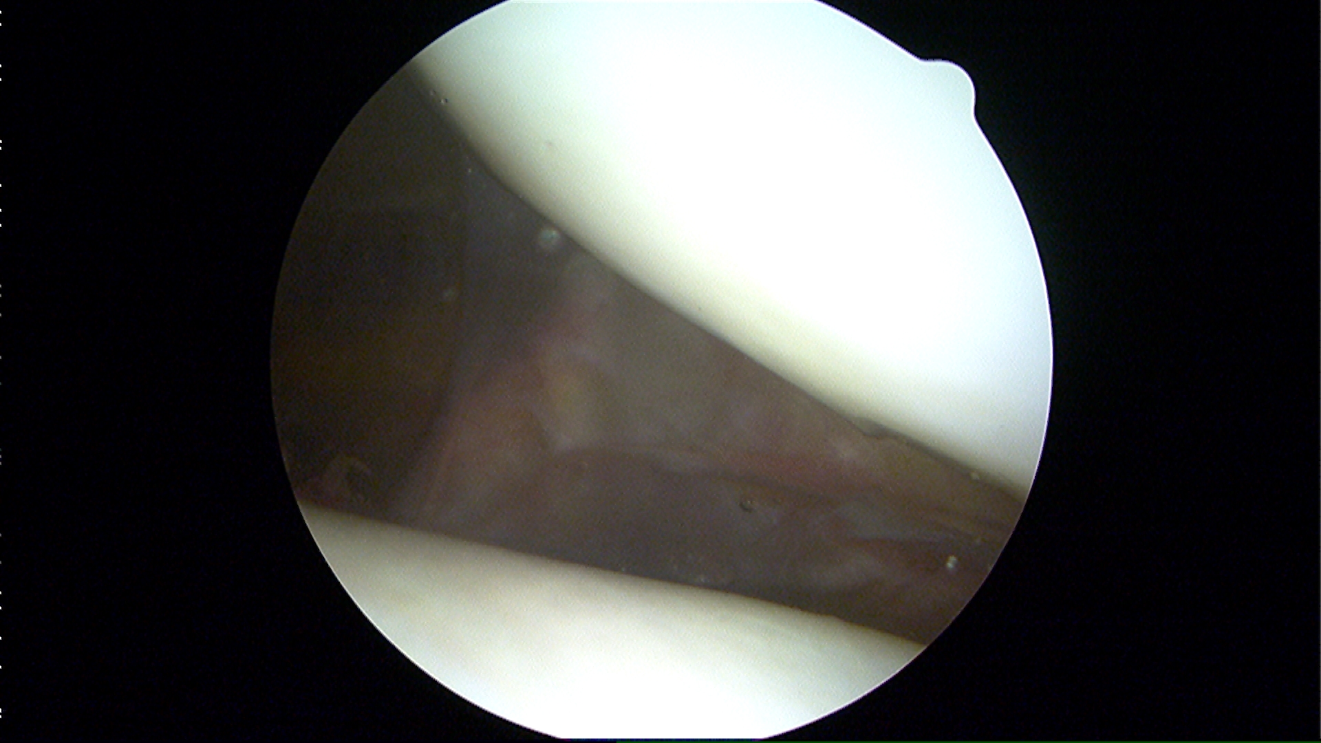
For those that understand physics, the coefficient of friction of healthy human cartilage is as low as 0.002 – unmatched by any artificial joint. The ultra-low friction surface can normally cope with tens of millions of loading cycles, even when subject to loads several times body weight (due to the addition of very powerful muscle forces around the joint), but under excessive load the structure can fail.
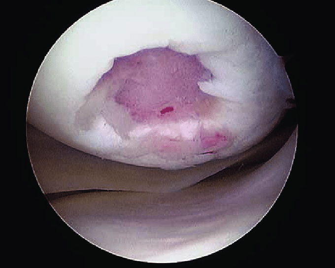
Damage to chondral surface cartilage can result in knee pain and swelling – pain because the underlying bone begins to take excessive loading stress which it doesn’t like, and swelling as a secondary reaction caused by inflammation. At a lower level of activity, this will often be tolerated, or prevented by reducing impact loading, but that is obviously not an option for Roger Federer whilst he hopes to continue his professional career.
There are various surgical options available for treating areas of surface cartilage damage. Once it becomes extensive, patients are usually faced with the prospect of joint replacement surgery, but if more isolated areas of damage are causing symptoms, then there are techniques that can be used to induce cartilage surface healing. These are generally more successful in younger patients who exhibit a better healing response (so even Roger at 40 may struggle with this).
The most simple of these goes back to the early days of orthopaedic surgery after WW2 when Ken Pridie, the first surgeon in Bristol to devote himself entirely to orthopaedics (and who also threw the discus and shotput in the Empire Games and was selected for the 1932 Olympics) developed a technique that came to be known as “Pridie drilling”.

He introduced the principle of creating multiple small drill holes in exposed bone (devoid of covering cartilage) with the objective of stimulating a fibrocartilage healing response. This type of technique is still in use today, performed via a keyhole surgery technique – either with a small sharp bone pick gently tapped into the surface (microfracturing) or with a thin drilling wire (microdrilling).
Microfracture surgery is suitable for cartilage defects with total area max 4cm2. There is a YouTube video of Microfracture – which shows how this is achieved. The stem cells that are released from the bone marrow by puncturing the exposed bone plate form mainly fibrocartilage (scar cartilage) – a process that takes several months to mature. It is a straightforward technique which gets good results if patients adhere to a strict regimen of crutch use, and high-repetition unloaded knee movements.
Larger areas are better treated with more complex surgical techniques that usually involve open incisions (rather than keyhole surgery):
- Autologous cartilage implantation (ACI) – using cells which are harvested from the knee, grown in a laboratory and then re-implanted at a second operation either under or within a patch of tissue to fill the defect – with an aim of produce hyaline-like cartilage which may last longer than fibrocartilage.
- Osteochondral autograft – moving plugs of bone and cartilage from areas of the knee that don’t take any direct load to the damaged weight-bearing zone.
- Osteochondral allograft – where larger areas are concerned, using donor bone and cartilage plugs to fill a defect.

Drawing of osteochondral grafting technique.
All of these operations require prolonged rehabilitation, and begin with a period of at least 6 weeks taking the load off the knee joint by using crutches, then a very gradual return to loading the joint. Federer says he will be “out of the game for many months”. I hope he makes it back as I still love watching him play.