
A ligament is a band of tough, flexible tissue made of collagen – a protein that forms long fibres. They can stretch a little under load, but if the force applied is too great, they can tear (or rupture).
There are 4 main ligaments that connect the thigh bone (femur) with the shin bones (tibia and fibula) that control how your knee is able to move:
- Medial Collateral Ligament (MCL) – a long strap-like ligament down the inner aspect of your knee which has 3 separate components, and also attaches to your inner cartilage pad (medial meniscus)
- Lateral Collateral Ligament (LCL) – a thick cord-shaped ligament from the outer femur to the fibula head
- Anterior and Posterior Cruciate Ligaments (ACL and PCL) – ligaments within the central section of your knee which cross over each other (hence cruciate, or crossing)

Ligaments of the knee.
There are also a variety of other smaller ligaments which act as fine balancers of the movement of this complex joint.
Knee ligament injuries are relatively common, typically resulting from awkward twisting movements during a slip or fall, or from direct impact such as in a rugby tackle. These ligaments have a plentiful nerve supply (hence severe pain at the time of injury), and also blood vessels within them (hence swelling and bleeding / bruising after injury). Patients will often feel a pop or a tear, and will normally be unable to continue activity (ie you can’t just “run it off”).
Medial Collateral Ligament – MCL
The MCL is the most commonly-injured knee ligament – either from a direct force applied to the outer aspect of the knee, or when the foot slips outwards and the knee collapses inwards. Most injuries are “sprains” – where the ligament fibres are injured but not completely disrupted (or only one of the 3 sections of the ligament is injured) – which are very painful initially but settle within 2-6 weeks (depending on the extent of the injury). There may be some localised swelling down the inner aspect of the knee, and some bruising may develop over the first few days. A simple short knee brace will help reduce pain in the early stages, and once the more acute pain has settled, an exercise programme guided by a physiotherapist is enough to return to full function.
More severe forces result in complete rupture or separation of the ligament. The knee will then feel unstable, like it is about to “give way”, and patients may feel the inner aspect of the knee joint (or medial compartment) opening up if they push the knee inwards. Surprisingly, once the ligament ends have separated, there may be less pain when compared to a less-serious partial injury (where any further movement pulls more on the damaged tissue). Even complete MCL tears can be treated without surgery, but it important that these are protected in a full (longer) hinged knee brace, initially with a restricted range of movement, so that they heal in the correct position. This would typically take around 6 weeks in a brace, with a return to sport at 8-10 weeks.
For a good video on MCL injuries and their treatment see Treatment & Exercises For MCL Strains on YouTube – it’s a popular video so you’ll have to get past a couple of adverts first.
Anterior Cruciate Ligament – ACL
Again commonly injured, the ACL sits within the cavity of the knee joint so when it ruptures and bleeds, the whole knee fills with blood, often within 10-20 minutes, giving rapid-onset swelling (and pain). Usually loaded during twisting or cutting movements, ACL rupture is a common (often non-contact) injury in football, netball and other similar sports. It is rarer to have a partial injury – once the fibres start to go, the process normally completes with both the bundles of the ligament completely separating.
Here is a video animation of the typical ACL injury mechanism: Anterior Cruciate Ligament (ACL) Injury – YouTube
Because the ligament is within the knee cavity, it is surrounded by the fluid that normally lubricates your joint, making it more difficult for the ends of the ruptured ligament to heal back together – so spontaneous healing of the ACL is much less likely than of the MCL.
Initial treatment starts with a physiotherapist – to overcome pain and swelling, to get the knee joint moving, and to work to prevent quads muscle wasting. Professional athletes will normally make an early decision to have operative reconstruction (usually at 2-3 weeks from injury), but most other patients will be encouraged to have 3-4 months of physiotherapy to maximise non-operative progress before deciding whether surgery is for them. It is however worth seeing an orthopaedic surgeon in the early weeks to check progress and to fully understand what factors might influence the decision to have reconstruction surgery.
We have a whole page on ACL surgery and post-operative rehab.
More information on how to reduce your risk of ACL rupture (and other knee injuries) can be found here: Avoiding knee injuries in sport.
Posterior Cruciate Ligament and Lateral Collateral Ligament – PCL and LCL
PCL and LCL injuries are much less common. They are both thick, strong ligaments that rarely come under enough force to cause rupture. The PCL can rupture with a fall from height directly onto the knee, or from the knee impacting a car dashboard in a front end vehicle collision. It can also be injured as part of a more complex pattern of multiple-ligament injury with higher levels of force, such as those sustained in a motorbike accident.
Early diagnosis for PCL injuries is key (requiring specialist examination and usually an MRI scan) as a specific type of knee brace is required to prevent the shinbone from sagging, and these usually get a good outcome without the need for surgical reconstruction.
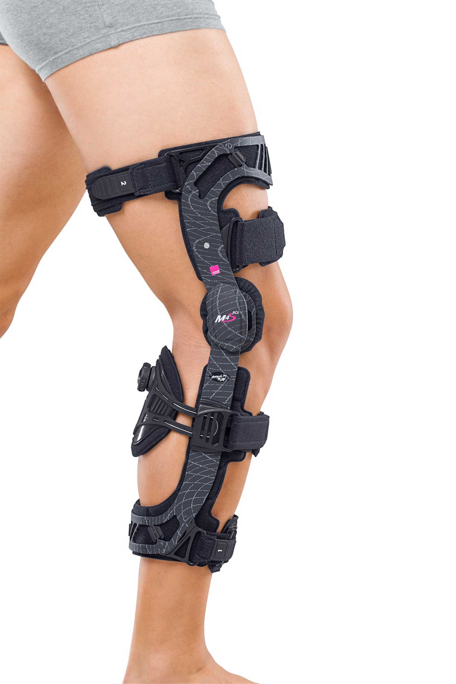
PCL Jack brace required to prevent sagging after PCL injury.
More complex patterns of multi-ligament knee injury are more likely to benefit from surgical reconstruction, and due to limited options to take appropriate graft tissue from the injured patient, surgeons will more often use human donor tissue (known as allograft) or artificial reconstruction ligaments, such as the synthetic “LARS” ligament seen in the image below, which act as a scaffold for human tissue to grow into.

Synthetic ligament used to re-inforce patient’s damaged tissue.